


Abstract
A comprehensive study of osteology remains a cornerstone of current orthopaedic and traumatological education. Osteology was already established as an important part of surgical education by the 16th century. In order to teach anatomy and osteology, the corpses of executed criminals were dissected by the praelector anatomiae of the Amsterdam Guild of Surgeons. Magnificent anatomical atlases preserve the knowledge obtained from these dissections. We present an overview of the most authoritative works of Vesalius, Bidloo, Cheselden, and Albinus authored in the 16th, 17th and 18th centuries. At that time a knowledge of osteology was necessary to pass the ‘master-exam’ in order to become a surgeon, and anatomical teaching was traditionally based on the practice of dissection. In the modern era, anatomical dissection and illustrations are largely being replaced by three-dimensional imaging and computer simulations, with an unfortunate trend in current curricula away from the established teaching technique of dissection. Education through the practice of dissection, particularly for future surgeons, remains integral to the development of tissue handling techniques, understanding of anatomical variation, and furthering of spatial awareness skills. With this review, we seek to remind contemporary surgeons of the lessons we can learn from our predecessors who valued education through anatomical dissection.
Introduction
‘Osteology’, derived from the Greek words osteon (bone) and logos (knowledge), is defined as the study of the structure and function of bones. The regulations of the Amsterdam Guild of Surgeons show that osteology was an important component of their surgical curriculum at the beginning of the 17th century.1,2 Historically, surgeons were trained under the guidance of a master-surgeon in a surgeon’s shop. In 1555, the Amsterdam Guild of Surgeons was granted the privilege of dissecting the bodies of executed criminals to teach anatomy.2 These lessons were given by the praelector anatomiae (lecturer in anatomy) in the Anatomy Theatre of Amsterdam. The praelector was entrusted with the education of the guild members.
The Osteology Lesson of Dr. Sebastiaen Egbertsz, 1619 belongs to the collection of paintings of the Amsterdam Guild of Surgeons and depicts one of the earliest lessons in osteology (Fig. 1).3 The painting records a carefully composed group portrait of the guild officials, which is reminiscent of the praelector’s osteology lessons. The skeleton serves as a central element in the composition and is thought to have been from an executed English pirate whose corpse was dissected in 1615. The painting clearly shows the removal of the skullcap with subsequent reattachment suggesting the purpose of the skeleton for teaching osteology.3
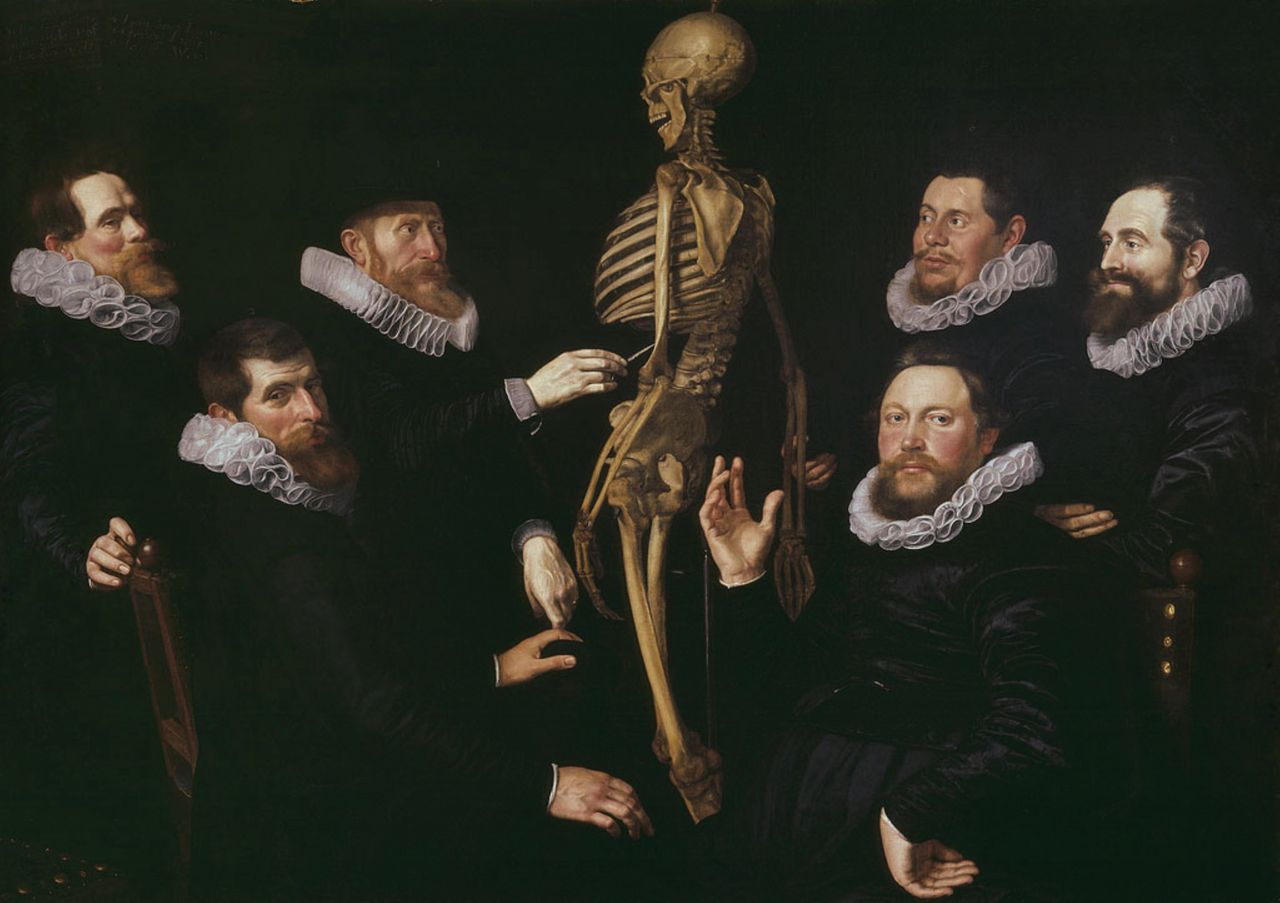
Fig. 1
TheOsteology Lesson of Dr. Sebastiaen Egbertsz, 1619 painted by Thomas de Keyser. Dr Sebastiaen Egbertsz (1563 to 1621) is standing on the left side of the skeleton and demonstrates the lower rib. Courtesy of the Amsterdam Museum.
We have reviewed original historical sources including atlases and exam reports contained in the comprehensive library collections held in Universities throughout The Netherlands. Taken together, these authoritative works on osteology provide a unique insight into the knowledge of leading physicians through the 16th to 18th centuries. Anatomical atlases from Vesalius, Bidloo, Cheselden, and Albinus demonstrate the knowledge of osteology provided by anatomical dissections,4-8 and exam transcripts from the master-exams (exams in surgical skills) reveal the emphasis placed upon osteology as a key component of surgical training.9-12 The ongoing accumulation of knowledge in this field resulted in the original 18th century surgical textbook of Heister and Ulhoorn, entitled Surgical Lessons.13 This book dominated the education of hundreds of contemporary surgeons trained in Europe, and reveals some unique insights into the application of osteology in the daily practice of surgeons from that era.
Anatomical atlases from the 16th to 18th centuries
During the Middle ages, the study of human anatomy was prohibited by the religious authorities and consequently anatomical knowledge was derived from dissections of animals and the traditional Greek and Roman writings of Hippocrates (470 to 400 BC) and Galen (130 to 200 AD).14 It was not until the Renaissance that investigation of the anatomy of the human body was tolerated and knowledge started to surpass that of antiquity.14
Andreas Vesalius’ (1514 to 1564) revolution in osteology
Andreas Vesalius studied medicine in Leuven, Paris, and Venice. He was appointed Professor of Surgery at the medical school in Padua in 1537.14 In 1543, Vesalius published the first edition of De humani corporis fabrica, describing the anatomy of the human body.8 This seminal work was considered ‘the greatest of all books written about human anatomy’ and for this outstanding achievement, he was granted nobility in 1556.15 This book heralded a new era in the understanding of human anatomy and was based upon his own anatomical dissections of the human body.
De humani corporis fabrica was published as a series of seven books. The first book comprised some 80 000 words and 140 illustrations describing all the bones of the human body. In the original work, Vesalius provided detailed accurate anatomical illustrations (Fig. 2).8 He described the technique for dissection of corpses of both adults and children, followed by the removal of soft-tissue remnants by boiling the bones. He recognised the importance of the study of the relationships between the bones, and developed a technique to reconstruct the complete skeleton using copper wire to articulate the individual bones.
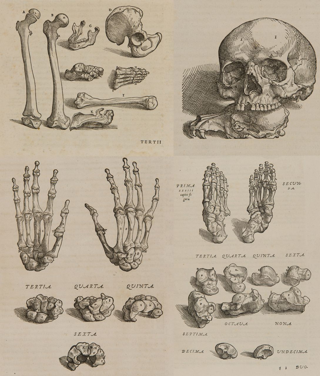
Fig. 2
Original anatomical illustrations of the osteology from Andreas Vesalius’ work entitledDe humani corporis fabrica libri primus (1555).8 Courtesy of University Library of Groningen, special collections (uklu KW C 569).
Vesalius successfully challenged several traditional concepts of osteology propagated by Galen. He identified the marrow cavity and postulated its importance in the nutrition of bone. He recognised the role of the epiphysis in growth, and emphasised the importance of cartilage connections between bones to maintain optimal joint function. From anatomical dissections of neonates in Bologna, Vesalius observed that the human skull had sutures, and even the smallest details of the auditory bones did not slip his attention. He discussed the morphology of bones and compared his observations with contemporary knowledge. In many respects Vesalius is the 16th century father of modern osteology.
Govard Bidloo (1649 to 1713) professional drawings
Govard Bidloo was Professor of Anatomy in The Hague (1688) and Leiden (1694).16His magnificent anatomical atlas, entitled Ontleding des Menschelijken Lichaams (Dissection of the human body), was published in Dutch in 1690.6 Bidloo praised the work of Vesalius and stated that his anatomical atlas was based on anatomical dissections, which were intended for educational purposes. Bidloo collaborated with a draftsman, Gérard De Lairesse (1640 to 1711), a pupil of Rembrandt and one of Amsterdam’s leading artists.17,18 Bidloo provided 105 accurate anatomical drawings to illustrate the volume.6 The sixth chapter, entitled ‘Whole osteology’, contained 16 drawings on osteology. However, in De Lairesse’s drawings an element of artistic licence can be seen. For example, one of the skeletons is holding a shroud over a grave, representing death itself (Fig. 3). While the artistic value of Bidloo’s atlas is recognised, its usefulness for surgeons has been disputed18 and the importance of this atlas is the successful collaboration between a physician and an artist.
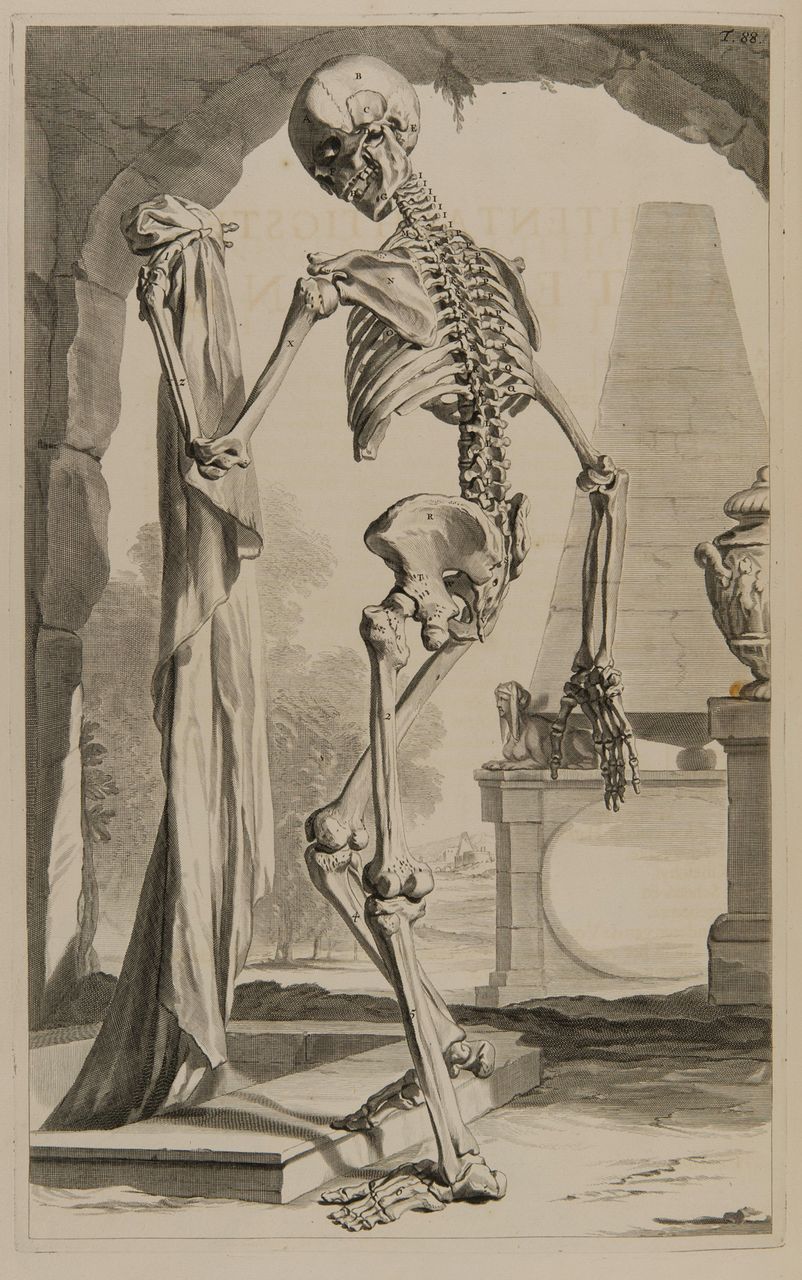
Fig. 3
Original anatomical drawing of the human skeleton by Gérard De Lairesse from Govard Bidloo’s work entitledOntleding des Menschelijken Lichaams (1690).6 The skeleton represents death itself by holding a shroud over a grave. Courtesy of University Library of Groningen, special collections (uklu KW C 1150).
William Cheselden (1688 to 1752) and the camera obscura
William Cheselden, one of the most prominent British surgeons and scientists of the 18th century,19 was introduced to the works of Vesalius and Bidloo through his teacher, William Cowper (1666 to 1709). Cheselden undertook his own anatomical dissections using the corpses of executed criminals to teach anatomy19 and osteology. His own anatomical atlas, entitled Osteographia, orthe Anatomy of the Bones (1733) is considered ‘one of the best English works on anatomy’ and merits a prominent place in the early series of anatomical atlases on osteology.7,19Osteographia contains 56 pages with anatomical illustrations of bones, ligaments, and cartilage structures.7 Cheselden was the first anatomist to make use of the camera obscura (pinhole camera) to prepare his anatomical illustrations. Unlike his predecessors, Cheselden included diseased specimens of bone in his anatomical atlas, and several illustrations demonstrating spinal pathologies. His atlas records the first examples of osteomyelitis in tuberculosis, the destruction of bone seen in advanced syphilis and some extreme forms of callus from untreated fractures (Fig. 4). Cheselden’s contribution was significant, having employed the technology of the camera obscura to accurately demonstrate and record the anatomy and pathology of bones to his students.
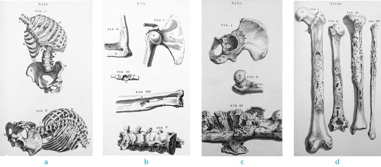
Fig. 4
Original anatomical illustrations of the osteology of William Cheselden’s work entitledOsteographia, or the Anatomy of the Bones (1733).7 The illustrations show in Figure 4a – Parts 1 & 2 : kyphoscoliosis of the spine. Figure 4b – Part 1: dislocation of the humeral head after a scapular fracture; Part 2: congenital ankylosis of the elbow; Part 3: osteomyelitis of the thumb in tuberculosis, Part 4: synostosis between the radius and ulna after an antebrachial fracture; Part 5: ankylosis of the lumbar spine. Figure 4c – Parts 1 & 2: destruction and perforation of the acetabulum by an abscess of the hip joint; Part 3: osteomyelitis of the femur. Figure 4d – Parts 1 to 4: destruction of bone in an advanced stage of syphilis. Authors’ collection.
Bernard Siegfried Albinus (1697 to 1770) homo perfectus
Bernard Siegfried Albinus was Professor of Anatomy and Surgery in Leiden. In 1747 he published his anatomical atlas Tabulae sceleti et musculorum, depicting what he termed the ‘homo perfectus’ (the ideal human).4 Albinus was convinced that an anatomist should understand the construction of the human body, as an architect must understand the foundations of his building.20 He considered the skeleton to be the foundation of the body and studied its anatomy to understand the mechanical functions of the skeleton and muscles. In 1753 Albinus collaborated with the artist Jan Wandelaar (1690 to 1759),20and published a further purely osteological anatomical atlas entitled Tabulae ossium humanorum.5 Albinus and Wandelaar developed a system to bring some objectivity to their depictions of the skeleton, which adopted wooden frames with grids of wires to achieve objectivity, symmetry, and vitality.20Tabulae ossium humanorum was published in large format (72 cm × 50 cm) and contained 34 anatomical illustrations on osteology.5 In each anatomical illustration the relationship between the bones and tendons, ligaments or joints were shown in intricate detail. The accuracy of their new system and their achievement is well demonstrated with Albinus’ fine depiction of the complex osteology of the foot (Fig. 5). Albinus was the first renaissance anatomist and surgeon to have insight into the mechanical functions of the body. His atlases record his ability to shed light on biomechanics, integrating the knowledge of osteology, with that of muscles and tendons.
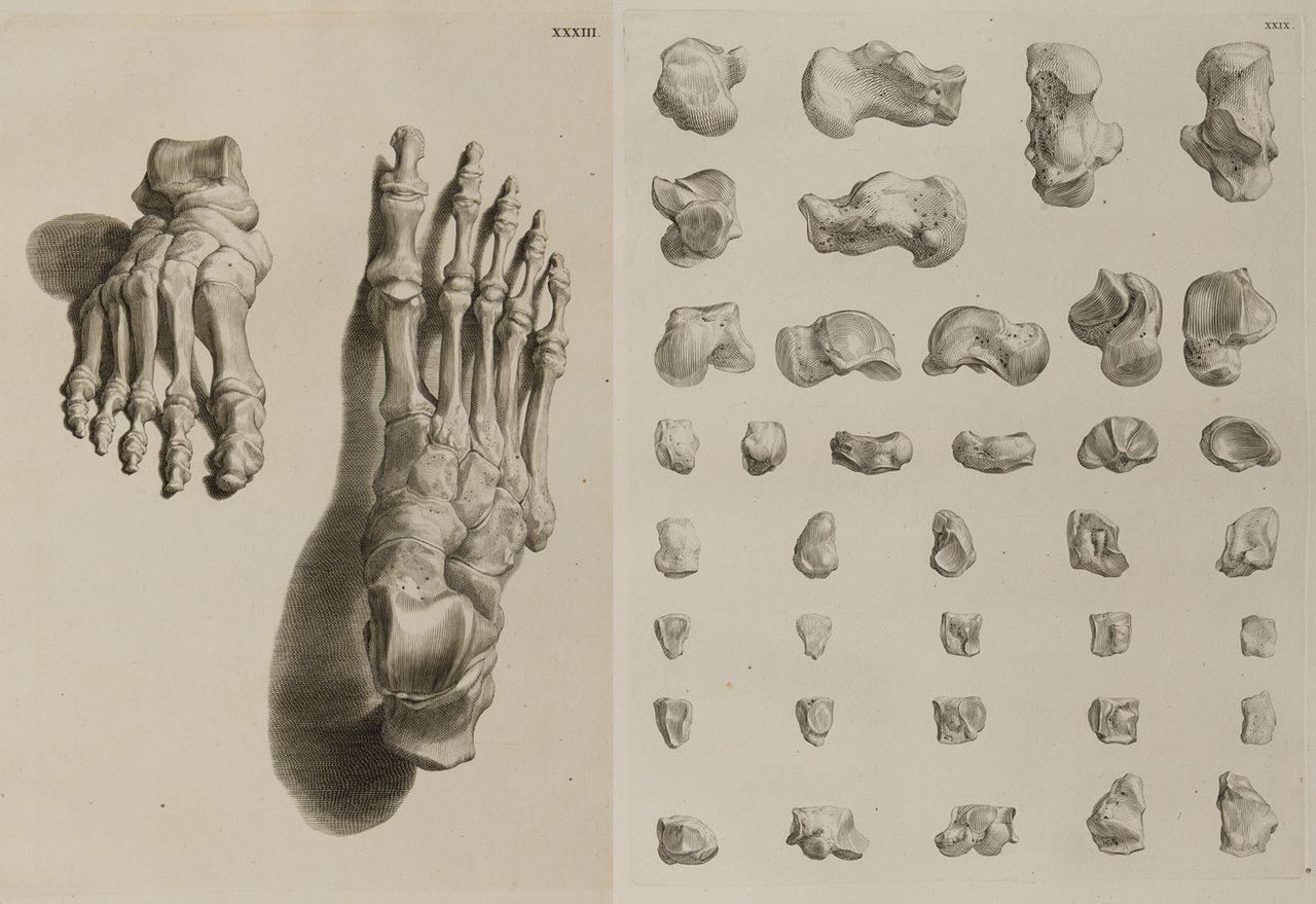
Fig. 5
Osteology of the foot, illustration from Bernard Siegfried Albinus’ work entitledTabulae ossium humanorum (1753).5 Courtesy of University Library of Groningen, special collections (uklu KW C 563).
Reports of the surgical master-exams
Knowledge of osteology was not confined to anatomical dissections and atlases. The training of surgeons from the 16th century onwards was completed by sitting and passing a master-exam.1 The exam was conducted by the board of the guild and the praelector anatomiae. The front piece of ‘t Nieuwe examen der chirurgie (1693) (The new exam in surgery) by Bernardus De Bout depicts an examinee under examination (Fig. 6).9 During the exam, the surgeons in training were required to both manufacture their own instruments and perform practical procedures. De Bout records that candidates were expected to manufacture their own lancets, and that these should be able to cut a piece of leather. The practical part of the examination included performing phlebotomies, the application of wound dressings and bandages. The master-exam was completed with a theoretical test and included knowledge of osteology and fractures. Similar to the current system of Royal College and Board exams, if the trainee passed his surgical exam he was granted the privilege to join the guild and to open his own surgeon’s shop in the city of his residence.

Fig. 6
Master-exam (examination in surgical training) in which the examinee is standing in front of the guild members. The cabinets with surgical instruments and osteology collection of the guild are clearly visible in the background of the drawing. FromtNieuwe examen der chirurgie, by De Bout (1693).9 Courtesy of University Library of Amsterdam, special collections (OTM: OK 61-1920 (1)).
The reports of the Dutch master-exams of the 17th and 18th centuries provide an insight into the importance of osteology in the training of surgeons, and carefully document discussions between trainee and master surgeons in viva voce style examinations.9-12 In remarkable similarities to current professional exams the master surgeon would ask questions about the anatomy and morphology of bones with examinees often responding with definition of the periosteum, diaphysis, apophysis, and epiphysis. Another favourite examination topic was the bony anatomy of the skull, including the finer details of the osteology and foraminae.
In one particularly well recorded examination a candidate was asked, ‘describe the anatomy of the vertebrae’. The candidate apparently replied, ‘They are all hollowed out as a ring through which the spinal cord runs, the cervical vertebrae are somewhat curved inwards (lordosis) and the thoracic vertebrae and sacrum are curved outwards (kyphosis)’. The vertebral body is flat, wide and half-round, and ‘from behind and sideways there are protrusions (spinous and transverse processes) of which the shape depends on the vertebral level’; answers that would not be too out of place in an examination today. Candidates were expected to have an in-depth knowledge of the topic, questions extending to the tiny ‘seed bones’ (sesamoids), which one candidate was keen to explain, ‘they grow between the tendons and are hidden underneath the ligaments’.
Heister’s and Ulhoorn’s ‘Heelkundige Onderwijzingen’ (Surgical Lessons) on osteology ‘applied’ in fracture treatment
Knowledge of osteology was not just a means of examination and a gateway to the professional body, it was also valuable for surgeons in their daily practice, most particularly in the treatment of fractures. ‘To understand a fracture and its healing, it is necessary to know the anatomy‘, was the opinion of the German and Dutch surgeons Laurens Heister (1683 to 1758) and Hendrik Ulhoorn (1687 to 1746).21 One of the most influential surgical textbooks in 18th century Europe was Heister’s work, simply entitled Chirurgie (Surgery).22,23 Ulhoorn translated the work into Dutch, but was liberal in his interpretation and added many of his own annotations, rebranding the Dutch title Heelkundige Onderwijzingen (Surgical Lessons) (1755).13
Heister and Ulhoorn outlined the process of fracture healing. They hypothesised that healing resulted from the leakage of fluid from the fractured bones. The fluid was released as the result of torn tubes in the bone, through which the ‘nourishing bone fluids’ circulate. The fluid bridged the fracture, and they called it ‘callus’.13
Fractures at this time were mainly treated conservatively by basic principles similar in concept to those utilised today; reduction and immobilisation using bandages or splints. To maintain reduction the authors usually advocated traction and described techniques using belts, ropes, and pulleys. Immobilisation was achieved with bandages and splints (typically made of wood, cardboard, copper, tin, or lead). It was not until the 19th century that dextrin and plaster were used to maintain immobilisation, and the forerunners of the modern ‘plaster of paris’ were strengthened bandages, achieved using egg-whites and soft wheat. Early attempts to intervene in fracture healing included removal of foreign bodies, bone fragments from open fractures, or even improving fracture healing through soaking of bandages in brandy, vinegar, rose oil, or herbal extracts.21 Occurring several hundred years prior to the invention of radiographs, surgeons relied on a sound knowledge of osteology to diagnose the type of fracture and to provide appropriate treatment for each specific fracture. Even today, many fractures are treated using the same basic principles.
Discussion
Anatomical education has long been, and still is, a cornerstone of surgical training. Osteology provided the foundation of orthopaedics and traumatology. The Osteology Lesson of Dr Sebastiaen Egbertsz, 1619 from the series of paintings of the Amsterdam Guild of Surgeons illustrates how important this education was in the earliest days of modern surgical practice. The image of the praelector anatomiae teaching human osteology to the guild members is present in the Guild collection (Fig. 1), and the master-exams in the 17th century demonstrate the importance of osteology and anatomy to early surgeons (Fig. 6). According to Ulhoorn and Heister, ‘surgeons must have knowledge of osteology for a rational treatment of fractures’.13 Their Surgical Lessons (1755) give us the opportunity to understand the practical methods used by surgeons in fracture treatment which have formed the foundation of modern orthopaedic and trauma practice.13
Early anatomical atlases were often produced through a collaboration between a physician and an artist.18 These anatomical illustrations evolved into a special category of art that contributed to medical science. In their drawings, the artists captured the findings of physicians’ anatomical dissections often with the allegory of mortality hidden in the illustrations, for example by a skeleton holding a shroud over a grave (Fig. 3).18 These old atlases of osteology are of outstanding quality in comparison with their modern counterparts. Cheselden’s atlas (1733) is distinct from others of this era in that it includes bone pathology notes along with drawings of pathological specimens of osteomyelitis, exostosis, and untreated fractures.7 Perhaps he was the first doctor to link pathology with abnormal morphology of the skeleton. During the 18th century the allegoric elements in the anatomical illustrations gradually disappeared.18 With time, the techniques for printing anatomical illustrations evolved from Vesalius’ wood engravings to copperplate engravings and eventually, to the use of lithography in the 19th century.18
Three-dimensional imaging technology was introduced, and has a growing role in anatomical teaching following the advent of CT and MR in the second half of the 20th century. TheVisible Human Project (1993) represents the ultimate attempt in three-dimensional imaging of the anatomy of the human body, including osteology.18 The body of a criminal, executed in Texas, was frozen and shaved into thin slices. Each cross-section was photographed and digitised. It is noteworthy how history repeats itself with the corpse of an executed criminal providing the material to teach anatomy.
In the era of the Surgeons’ Guild, surgical training comprised an apprenticeship in a surgeon’s shop with education in the anatomical theatre. From these early origins, surgical training has evolved into an academic, structured medical curriculum followed by specialisation in surgery. The teaching of anatomy, including lessons in osteology, has changed substantially over the past centuries.24 Anatomical dissections, using the corpses of executed criminals, were recorded in magnificent anatomical atlases over more than 400 years ago, and formed the basis for the anatomical education in this period.
More recently, the introduction to medical school syllabuses of digital anatomical imaging is allowing students to perform virtual dissections and has reduced the need for cadaveric dissection. Whilst having many obvious advantages, including gaining familiarity with modern imaging techniques in some curricula, students are less frequently exposed to anatomical dissections.24-26 Great care must be taken as these technologies develop to ensure there remains a place for performing dissection which not only teaches anatomy but also helps to develop sufficient understanding of human tissues, spatial aptitude and, anatomical variations.24-26 Our forefathers like Vesalius, Bidloo, Cheselden, Albinus, Heister, and Ulhoorn all taught direct experience using human material for anatomical learning. We must strive in the modern era of teaching and learning to capitalise on the lessons of our forefathers who, over 400 years ago, provided sufficient learning materials to allow surgeons to acquire substantial knowledge of osteology from anatomical dissections. This tried and tested approach should remain a keystone for the continued training and examination of surgeons.
1 IJpma FF , van de GraafRC, PierikEG, van GulikTM. De meesterproef in de chirurgijnsopleiding. Ned Tijdschr Geneeskd2010;154:A795. Google Scholar
2 Meyer H. Privilegien, willekeuren en ordonnantiën, betreffende het Collegium Chirurgicum Amstelaedamense. Amsterdam: Pieter van den Berge, 1736. Google Scholar
3 Middelkoop N, Noble P, Wadum J, Broos B. Rembrandt under the scalpel. The anatomy lesson of Dr Nicolaes Tulp dissected. Amsterdam: Six Art Promotion, 1998. Google Scholar
4 Albini BS. Tabulae sceleti et musculorum corporis humani. Leiden: Lugduni Batavorum, prostant apud Joannem & Hermannum Verbeek, 1747. Google Scholar
5 Albini BS. Tabulae ossium humanorum. Leiden: Joannem & Hermannum Verbeek, 1753. Google Scholar
6 Bidloo G. Ontleding des menschelijken lichaams. Uitgebeeld, naar het leven in honderd en vyf aftekeningen door de Heer Gerard de Lairesse. Amsterdam: De weduwe van Joannes van Someren, de erfgenamen Joannes van Dijk, Hendrik en de weduwe Dirk Boom, 1690. Google Scholar
7 Cheselden W. Osteographia, or the Anatomy of the Bones. London: William Bowyer, 1733. Google Scholar
8 Vesalius A. De humani corporis fabrica libri primus. Basileae: Ioannem Oporinum, 1555. Google Scholar
9 De Bout B. ’t Nieuwe examen der chirurgie. Amsterdam: Jan ten Hoorn, 1693. Google Scholar
10 Herls C. Examen der Chyrurgie. Middelburgh: Françoys Kroock, 1663. Google Scholar
11 Rhijnenburgh BI. Examen ofte proeve der chirurgijns ende barbieren. Rotterdam: Pieter van Waesberge, 1650. Google Scholar
12 Verbrugge J. Heel-konstige examen ofte instructie der chirurgie begrepen in vier tractaten. Amsterdam: Jan Claesz Ten Hoorn, 1677. Google Scholar
13 Ulhoorn H. Laurens Heisters heelkundige onderwijzingen. Amsterdam: Gerrit de Groot en Jan Morterre, 1755. Google Scholar
14 Baruch JZ. Leven en werk van Andreas Vesalius. Gravenhage: Kruseman’s, 1964. Google Scholar
15 Richardson WF, Carman JB. On the Fabric of the Human Body: A Translation of De Humani Corporis Fabrica Libri Septem. Book I: the Bones and Cartilages. Andreas Vesalius. San Francisco: Norman publishing, 1998. Google Scholar
16 Lindeboom GA . Cowper’s brutale ‘plagiaat’ van Bidloo’s anatomische atlas. Ned Tijdschr Geneeskd1982;126:1878–1882. Google Scholar
17 Molenaar JC . Anatomie als schouwspel. Uit de bibliotheek van de Vereniging Nederlands Tijdschrift voor Geneeskunde. Govard Bidloo: Ontleding des Menschelijken Lichaams; 1689 en William Cowper: The Anatomy of Humane Bodies; 1698. :. Ned Tijdschr Geneeskd2004;148:2594–2602. Google Scholar
18 Rifkin BA, Ackerman MJ. Menselijke anatomie, van de renaissance tot het digitale tijdperk. Amsterdam: Mets & Schilt, 2006. Google Scholar
19 Sanders MA . William Cheselden: anatomist, surgeon, and medical illustrator. Spine1999;24:2282–2289.CrossrefPubMed Google Scholar
20 Hildebrand R . Attic perfection in anatomy: Bernhard Siegfried Albinus (1697-1770) and Samuel Thomas Soemmerring (1755-1830). Ann Anat2005;187:555–573.CrossrefPubMed Google Scholar
21 Van Loon L. Historisch overzicht van de fractuurbehandeling der lange pijpbeenderen. Wageningen: N.V. Gebr. Zomer & Keuning’s uitgevers Mij, 1935. Google Scholar
22 Haeger K. The illustrated history of surgery. Gothenburg: AB Nordbok, 1988. Google Scholar
23 Heister L. Chirurgie. Nürnberg: Hoffmann, 1719. Google Scholar
24 Sugand K , AbrahamsP, KhuranaA. The anatomy of anatomy: a review for its modernization. Anat Sci Ed2010;3:83–93.CrossrefPubMed Google Scholar
25 DeFriez CB , MortonDA, HorwitzDS, et al.Orthopedic resident anatomy review course: a collaboration between anatomists and orthopaedic surgeons. Anat Sci Educ2011;4:285–293. Google Scholar
26 Gillingwater TH . The importance of exposure to human material in anatomical education: a philosophical perspective. Anat Sci Educ2008;1:264–266.CrossrefPubMed Google Scholar
